New Fund Established to Stimulate Regenerative Medicine Industry
MONTREAL, QUEBEC--(Marketwire -04/30/12)- With a shared goal of supporting development projects that will boost innovation in the growing field of stem cells and biomaterials-based products, Pfizer Canada and the Centre for Commercialization of Regenerative Medicine (CCRM) have established the Pfizer-CCRM Innovation Fund to accelerate regenerative medicine (RM) technologies for drug screening and therapeutic applications. The announcement is being made at the first annual Till & McCulloch Meetings (April 30-May 2), Canada's premier stem cell meeting, jointly hosted by the Stem Cell Network and CCRM.
"CCRM was created on the premise that it would work with academia and industry on projects that will hopefully move RM technologies and innovations from the bench to the bedside," says Michael May, CEO of CCRM. "Canada is already a leader in this field and additional funding to advance novel research through early product development will only make us stronger. We're very pleased to be partnering with Pfizer Canada and appreciate their confidence in joining with us."
"Pfizer Canada is pleased to contribute to this new fund which will support important research here in Canada," explains Dr. Bernard Prigent, Vice-President and Medical Director, Pfizer Canada. "With the novel resources offered through CCRM's development capabilities, we hope to help advance the RM field in this country."
Pfizer Canada has contributed a total of $500,000 to the Pfizer-CCRM Innovation Fund and CCRM will contribute matching dollars to any approved projects undertaken in the duration of this fund.
About Centre for Commercialization of Regenerative Medicine (CCRM)
CCRM, a Canadian not-for-profit organization funded by the Government of Canada's Networks of Centres of Excellence program and six institutional partners, supports the development of technologies that accelerate the commercialization of stem cell- and biomaterials-based technologies and therapies. A network of academics, industry and entrepreneurs, CCRM translates scientific discoveries into marketable products for patients. CCRM launched in Toronto's Discovery District on June 14, 2011.
About Pfizer Canada
Pfizer Canada Inc. is the Canadian operation of Pfizer Inc., the world's leading biopharmaceutical company. Pfizer discovers, develops, manufactures and markets prescription medicines for humans and animals. Pfizer Inc. invests more than US$7 billion annually in R&D to discover and develop innovative life-saving and life-enhancing medicines in a wide range of therapeutic areas. Our diversified health care portfolio includes human and animal biologic and small molecule medicines and vaccines, as well as nutritional products and many of the world's best-known consumer products. For more information, visit http://www.pfizer.ca
Read this article:
New Fund Established to Stimulate Regenerative Medicine Industry
Recommendation and review posted by sam
South Korea steps up stem-cell work
South Korea's government is focused on boosting funds for stem-cell research this year.
Jo Yong Hak/Reuters
The South Korean health ministry announced last month that research into stem cells and regenerative medicine will receive a funding boost of 33 billion won (US$29 million) in 2012, four times that given in 2011. Overall, six different ministries will invest 100 billion won in stem-cell research this year.
Until last year, public investment in stem cells in South Korea was relatively low and targeted mainly at basic research. But the country's Ministry of Health and Welfare is now expanding its support for clinical research on stem cells, with the money being used to link basic research to intermediate or clinical studies. The aim is to commercialize the research at an early stage.
"From the current research atmosphere and infrastructure, the government has judged that stem-cell studies are now maturing," says Hyung Min Chung, president of Seoul-based biotechnology firm Cha Bio and Diostech and an adviser on the budget plan. He adds that his company is particularly pleased that government investment decisions on developing stem-cell therapies will be made more quickly.
The government money will be allocated to two areas: rare or incurable diseases for which there is little incentive for private investment, such as spinal cord damage; and common chronic conditions, such as arthritis, for which the aim is to help South Korean companies to capture part of the large potential market for treatments.
South Korea has been trying to restore its tarnished image in the field after the fall of one of its stellar stem-cell scientists, Woo Suk Hwang, who was convicted in 2009 for embezzlement and bioethics violations after falsifying results. The country has begun to regain confidence from a series of successful research projects, and is ranked in the world's top ten for stem-cell and regenerative medicine research in terms of number and quality of publications. The South Korean government thinks that the country can be a top global competitor if the field is given enough support.
In the past year, the South Korean Food and Drug Administration (FDA) has approved the worlds first three stem-cell treatments Hearticellgram-AMI, Cupistem and Cartistem which followed on the heels of clinical tests for human embryonic stem-cell therapies approved in 2010, according to the health ministry.
But many are still wary of those therapies. Clearly, there has been increased international focus on what is going on in the Korean regenerative industry since the approval of three stem-cell products in the past year, which I think caught many industry observers off guard, says Douglas Sipp, who works on stem-cell policy and ethics at the RIKEN Center for Developmental Biology in Kobe, Japan. "It appears that the Korean FDA has been focusing on fast-tracking products through the approval pathway, he says, adding that it will be interesting to see what other regulatory authorities think about these products.
Dong Wook Han, a professor of stem-cell biology at Konkuk University in Seoul, is worried about how the country regulates clinical research on stem cells. "Compared to a country like Germany, where I studied and where the regulations on embryonic stem cells are much stricter, South Korea has more freedom in regulation, which means it can explore more opportunities in clinical research. However, he emphasizes that instead of moving too quickly, South Korea needs to adopt the stricter regulations from other countries to test safety and efficacy.
Read the original post:
South Korea steps up stem-cell work
Recommendation and review posted by sam
Duke Team Turns Scar Tissue into Heart Muscle Without Using Stem Cells
By Duke Medicine News and Communications
Scientists at Duke University Medical Center have shown the ability to turn scar tissue that forms after a heart attack into heart muscle cells using a new process that eliminates the need for stem cell transplant.
The study, published online April 26 in the journal Circulation Research, used molecules called microRNAs to trigger the cardiac tissue conversion in a lab dish and, for the first time, in a living mouse, demonstrating the potential of a simpler process for tissue regeneration.
If additional studies confirm the approach in human cells, it could lead to a new way for treating many of the 23 million people worldwide who suffer heart failure, which is often caused by scar tissue that develops after a heart attack. The approach could also have benefit beyond heart disease.
"This is a significant finding with many therapeutic implications," said Victor J. Dzau, MD, a senior author on the study who is James B. Duke professor of medicine and chancellor of health affairs at Duke University. "If you can do this in the heart, you can do it in the brain, the kidneys, and other tissues. This is a whole new way of regenerating tissue."
To initiate the regeneration, Dzau's team at Duke used microRNAs, which are molecules that serve as master regulators controlling the activity of multiple genes. Tailored in a specific combination, the microRNAs were delivered into scar tissue cells called fibroblasts, which develop after a heart attack and impair the organ's ability to pump blood.
Once deployed, the microRNAs reprogrammed fibroblasts to become cells resembling the cardiomyocytes that make up heart muscle. The Duke team not only proved this concept in the laboratory, but also demonstrated that the cell conversion could occur inside the body of a mouse -- a major requirement for regenerative medicine to become a potential therapy.
"This is one of the exciting things about our study," said Maria Mirotsou, PhD, assistant professor of cardiology at Duke and a senior author of the study. "We were able to achieve this tissue conversion in the heart with these microRNAs, which may be more practical for direct delivery into cells and allow for possible development of therapies without using genetic methods or transplantation of stem cells."
The researchers said using microRNA for tissue regeneration has several potential advantages over genetic methods or transplantation of stem cells, which have been difficult to manage inside the body. Notably, the microRNA process eliminates technical problems such as genetic alterations, while also avoiding the ethical dilemmas posed by stem cells.
"It's an exciting stage for reprogramming science," said Tilanthi M. Jayawardena, PhD, first author of the study. "It's a very young field, and we're all learning what it means to switch a cell's fate. We believe we've uncovered a way for it to be done, and that it has a lot of potential."
Go here to see the original:
Duke Team Turns Scar Tissue into Heart Muscle Without Using Stem Cells
Recommendation and review posted by Bethany Smith
Fat Grafting with Concentrated Stemcells vs. Traditional Facelift
Fort Lauderdale, FL (PRWEB) April 29, 2012
Dr. Sassani states that he has been achieving significant and natural looking results with the addition of fat grafting with concentrated stem-cells to the list of services that he provides. The ideal candidate for this new innovative procedure has suffered a loss of volume of the face, which compromises skin texture. The skin may appear thinner lacking its once youthful volume.
Though injectables have their place in anti-aging, there is a significant difference between fat grafting with concentrated stem cells and all other injectible procedures. All injectables such as, Botox, Juvederm and Radiesse are composed of synthetic materials. In contrast, fat grafting with concentrated stem cell is completely natural because it is composed 100% of the individuals own fat cells. Fat grafting with concentrated stem cells offers something no other injectable can; use of the patients own tissue.
The fat is harvested from the lower abdomen and/or inner thighs to provide the volume necessary to correct the deficiency in the face. As an added bonus, the stem-cells that are transported along with the fat grafts are believed to improve overall skin color, texture and pore size. Stem cells are the repair cells of the body as they morph to become whatever cells they come in contact with. The harvested fat is spun in a centrifuge to concentrate these stem-cells for injection.
When should one have a facelift and when should you consider fat grafting with concentrated stem cells?
This procedure can be done either alone or in conjunction with facial aesthetic procedures. For patients exhibiting a loss of facial volume only, fat grafting with concentrated stem-cells done alone can be the ideal solution.
As the name suggests, facelifts lift sagging skin, tone the face and neck muscles, and remove the wrinkles. The ideal candidate for a facelift has suffered a loss of skin elasticity and muscle tone of the face and neck. Often the candidate will appear tired.
For candidates desiring a lift and tone of the face and neck with volume restoration, a facelift in combination with fat grafting may be the best solution. This candidate will have suffered a loss of skin elasticity, muscle tone and noticeable depletion of volume to the face.
The recovery time for all the procedures is approximately two weeks. After this time, the patient may comfortably return to normal social activities and interaction. A patients skin quality, texture, and tone will continue to improve as the stem-cells integrate. The end result is a tailored and youthful appearance with volume restoration and the skin noticeably improved overall. Patients will appreciate that the rejuvenative effects are enduring.
Take Shape Plastic Surgery, PA offers free consultations. They welcome you to visit their Medicare-certified, state-licensed, nationally-accredited, ambulatory surgical center in Fort Lauderdale, FL. You can check out their website at http://www.takeshape.info or call (954)585-3800 to schedule an appointment.
Read the original:
Fat Grafting with Concentrated Stemcells vs. Traditional Facelift
Recommendation and review posted by Bethany Smith
March of Dimes awards $250,000 prize to 2 scientists who pioneered advances in skin disorders
Public release date: 30-Apr-2012 [ | E-mail | Share ]
Contact: Elizabeth Lynch elynch@marchofdimes.com 914-997-4286 March of Dimes Foundation
WHITE PLAINS, NY April 30, 2012 Two scientists who have revolutionized the understanding of skin biology, creating crucial advances in treating skin cancers and other diseases as well as severe burns, have been chosen to receive the 2012 March of Dimes Prize in Developmental Biology.
Howard Green, MD, George Higginson Professor of Cell Biology, Department of Cell Biology, Harvard Medical School, and Elaine Fuchs, PhD, Rebecca C. Lancefield Professor, Laboratory of Mammalian Cell Biology & Development, Howard Hughes Medical Investigator, Rockefeller University, will share this year's prize.
Their work pioneered innovative technologies that explain the molecular underpinnigns of skin stem cells and inherited skin disorders, including cancers and some birth defects.
"Taken together, the research of Dr. Green and Dr. Fuchs has expanded medicine's ability to diagnose and understand the basis of many skin disorders, from cancer to inherited disorders to severe burns," said Joseph Leigh Simpson, MD, senior vice president for Research and Global Programs at the March of Dimes. "Their work has saved the lives of thousands of burn patients and we hope their work with skin stem cells will lead to new ways to prevent and treat birth defects."
Dr. Green and Dr. Fuchs have worked closely together. Dr. Fuchs is a former postdoctoral fellow in Dr. Green's lab, and throughout their careers they shared their scientific findings. Many of their findings have been translated into specific treatments.
Dr. Fuchs uncovered the genetic basis of blistering skin diseases and deciphered the characteristics of skin stem cells that allow them to develop into distinct tissues and organs. She also pioneered the use of reverse genetics, which studies protein functions and then determines what diseases occur when the proteins malfunction. She studies how skin stem cells become activated to form hair and skin, how they adapt to heal wounds, and how mutations in the activation process can lead to skin cancers, such as pilomatrichomas and squamous cell carcinomas. Her pioneering work holds promise for possible therapies for baldness.
Dr. Green, considered a founding father of regenerative medicine, developed a permanent skin restoration treatment that has saved the lives of thousands of burn victims. He previously developed the first therapeutic use of cells grown in a lab. His worked laid the foundation for the discovery of genes that are responsible for genetic skin defects. He discovered that hybrid human and mouse cells lost their human chromosomes, leading to the methods used today for gene mapping.
Dr. Green and Dr. Fuchs will receive the Prize at a gala black-tie dinner and ceremony at the Fairmont Copley Plaza. They will deliver the Seventeenth Annual March of Dimes Prize Lectures at the Hynes Convention Center during the Pediatric Academic Societies annual meeting. CBS sportscaster Greg Gumbel, member of the March of Dimes national Honorary Board of Trustees, will host the award ceremony.
More here:
March of Dimes awards $250,000 prize to 2 scientists who pioneered advances in skin disorders
Recommendation and review posted by Bethany Smith
Before the Procedure – Stem Cell Therapy (IPO) – Video
28-04-2012 19:32 IPO was diagnosed with a partial tear of her cranial cruciate ligament through x-rays. I decided to try and treat her conservatively but ultimately decided to perform stem cell therapy on her since the procedure yields many positive results. This is the "Before" video... stay tuned in 3-4 weeks for updates on her results.
Read more from the original source:
Before the Procedure - Stem Cell Therapy (IPO) - Video
Recommendation and review posted by Bethany Smith
Stem cell therapy for WCMS student has remarkable results
When Tyler was born, the umbilical cord was wrapped around his neck, causing a lack of oxygen to his brain that led to Tyler suffering a stroke during delivery. The stroke caused damage to the back of Tylers brain. Tyler was diagnosed with cerebral palsy and his mother, Lisa Biermann, was told to expect the worst: a child who would never walk, talk, or have any chance at a normal life.
Lisa refused to give up hope. She tried everything she could to help Tyler. Tyler could not walk because his feet would not sit flat on the floor. She tried botox injections every three months, braces, casts and even ankle cord surgery. Nothing worked.
Lisa said Tyler could not communicate with her at all. She never knew when he was in pain because he was unable to tell her.
Tyler was considered to be blind, with a prescription that was over nine units nearsighted, and his eyes jumped around. Even with glasses, he could not focus his vision, and doctors did not believe he could see, or ever would see.
Until he was 8 years old, Lisa would carry Tyler from his classes at Woodland Park Elementary.
When Tyler was 8, he had a seizure. Dr. David Steenblock, who is based in California, heard about Tyler and offered to help him with umbilical cord stem cell therapy. Lisa said she thought hard about it, and because she had tried everything else and nothing had worked, she decided to try the stem cell therapy, which Dr. Steenblock told her had no side effects.
In December 2007, Lisa, Dr. Steenblock and his team took Tyler for the treatment, which had to be done in Tijuana, Mexico, because stem cells injection is currently not legal in the United States. Three months later, they went for a second injection.
The stem cells were given to Tyler intravenously for a period of approximately 45 minutes.
Lisa said within weeks, she saw monumental changes in Tyler. All the milestones he never reached as a baby, he began reaching.
Within three months Tyler could put his feet flat on the floor and could walk independently. At six months post-treatment, he no longer needed the painful braces that gave him bunions.
See the original post here:
Stem cell therapy for WCMS student has remarkable results
Recommendation and review posted by Bethany Smith
How stem cell therapy can keep the immune system under control
A new study, appearing in Cell Stem Cell and led by researchers at the University of Southern California, outlines the specifics of how autoimmune disorders can be controlled by infusions of mesenchymal stem cells.
Mesenchymal stem cells (MSC) are highly versatile stem cells that originate from the mesoderm, or middle layer of tissue, in a developing embryo. MSC can be isolated from many different kinds of human tissue, including bone marrow and the umbilical cord.
Principal investigator Songtao Shi, professor at the Ostrow School of Dentistry of USC Center for Craniofacial Molecular Biology, said that recent studies have shown the benefits of administering MSC to patients with immune-related disorders such as graft versus host disease, systemic lupus erythematosus, rheumatoid arthritis, and more.
These studies showed that infusions of MSC appeared to quell the production and function of overactive immune cells, including T- and B-lymphocytes. However, the specific mechanism behind how MSC get the immune cells under control hasn't been fully understood.
"Mesenchymal-Stem-Cell-Induced Immunoregulation Involves FAS-Ligand-/FAS-Mediated T Cell Apoptosis" shines light on how infused MSCs target and defeat overactive immune cells.
Examining the effects of MSC infusion in mice with systemic sclerosis (SS)-like immune disorders, Shi and his colleagues discovered that a specific cellular mechanism known as the FAS/FAS-ligand pathway was the key to the remarkable immune system benefits.
Specifically, in mice with SS-like disorders, infusions of MSC caused T-lymphocyte death with FASL/FAS signaling and lessened symptoms of the immune disorder. However, MSC deficient in FAS-ligand failed to treat immune disorders in SS-afflicted mice.
With the hopeful results of the animal model study in mind, Shi's colleagues in China performed a pilot study with patients suffering from systemic sclerosis. Infusions of MSCs provided similar clinical benefits to patients, and experimental analysis revealed that the FASL/FAS pathway was also at work in humans with SS.
The identification of the cellular workings responsible for the stem cell treatments' success may eventually help doctors find optimal cell-based treatment for some immune diseases, Shi said.
Basic research portions of this study were supported by the National Institute of Dental and Craniofacial Research and the California Institute for Regenerative Medicine. Clinical studies were supported by a grant from the China Major International (Regional) Joint Research Project.
See the original post here:
How stem cell therapy can keep the immune system under control
Recommendation and review posted by Bethany Smith
World Government Election Fraud 2012 Part 4 – Video
28-04-2012 12:02 Playlist: I work a job and don't make a lot of money at work. Please be patient for the other parts. I hope to complete this documentary before June 2012. This educational documentary World Government Election Fraud 2012 reveals research about government, politics, energy, sovereignty, sustainability and many other topics including solutions. Diebold sells computer voting machines they had Republican conflict of interest during the 2 term voting Fraud of George Bush. Ron Paul Admits he is running the same platform George Bush used to win in 2000! Be your own leader. Do your own Research. Ron Paul wrote a book called The Case for Gold with Lewis Lehrman. Lew is a papal knight (Sovereign Military Order of Malta) Lew was on the board of directors of The Project for a New American Century. Lewis Lehrman is a member of the CFR and The Council for National Policy. The CNP consists of powerful individuals in the military industrial complex (Black Water aka Xe) banking cartels, multi-national corporations, previous presidents/presidential candidates, Prime Minister of Canada, Evangelical Christian Zionists, Scientology, Mormons and a lot of the guests Alex Jones has on his radio "program" on a regular basis. Meaning that, it is behind the phony patriot movement, tea party, libertarians and other types of activism. Butcher of Russia, Vladimir Lenin, was a Freemason. Aleister Crowley (1875-1947) became a Freemason of the 33rd degree in the Scottish rite and was a ...
See the article here:
World Government Election Fraud 2012 Part 4 - Video
Recommendation and review posted by Bethany Smith
USP9x: Gene discovered can 'turn off' pancreatic cancer
Aggressive pancreatic tumors may be treatable with a new class of drugs because of the discovery of the USP9x gene, according to Cancer Research UK.
Less than one in five people with pancreatic cancer survive past the first year after being diagnosed, reported BBC News. The study, published in the journal Nature, showed that the gene in question was being switched off in cancerous cells. Drugs with the potential to turn USP9x back on and stop the spread of cancer are already being tested.
Research was first conducted on mice and then on human cell lines, according to AAP. In both cases, it showed that the gene is switched off by chemical "tags" on the surface of its DNA.
"We looked in human tumor specimens and we found that [USP9x] was missing in a fraction of patients the patients that did very poorly... the people who died the fastest," said researcher David Tuveson to News24. "Patients that had a low level of the gene expressed... they died very quickly after their operation and the patients who at the end of their life had lots of metastasis [spreading of the cancer], they had also a very low level of this protein."
More from GlobalPost:Cancer 'encyclopedia' to help personalize cancer care, scientists say
http://www.globalpost.com/dispatch/news/health/120430/usp9x-gene-discovered-pancreatic-cancer
Original post:
USP9x: Gene discovered can 'turn off' pancreatic cancer
Recommendation and review posted by Bethany Smith
Pancreatic cancer gene discovery
30 April 2012 Last updated at 02:45 ET
Aggressive pancreatic tumours may be treatable with a new class of drugs, according to Cancer Research UK
Less than one in five people with this form of cancer are still alive a year after being diagnosed.
A study, published in the journal Nature, showed that a gene was being switched off in the cancerous cells.
The reseachers said drugs were already being tested which had the potential to turn the gene back on, to stop the spread of the cancer.
Around 7,800 people in the UK are diagnosed with pancreatic cancer every year and it is the fifth most deadly cancer.
These results raise the possibility that a class of promising new cancer drugs may be effective at treating some pancreatic cancers
Studies in mice showed that a gene called USP9x, which normally stops a cell from dividing uncontrollably, is switched off in some pancreatic cancer cells.
The gene is not mutated, but other proteins and chemicals become stuck to it and turn the gene off.
Studies then showed that UPS9x was being turned off in human pancreatic cancer.
The rest is here:
Pancreatic cancer gene discovery
Recommendation and review posted by Bethany Smith
Improved Adult-Derived Human Stem Cells Have Fewer Genetic Changes Than Expected
--Study lends support to safe use for therapy
Newswise A team of researchers from Johns Hopkins University and the National Human Genome Research Institute has evaluated the whole genomic sequence of stem cells derived from human bone marrow cellsso-called induced pluripotent stem (iPS) cellsand found that relatively few genetic changes occur during stem cell conversion by an improved method. The findings, reported in the March issue of Cell Stem Cell, the official journal of the International Society for Stem Cell Research (ISSCR), will be presented at the annual ISSCR meeting in June.
Our results show that human iPS cells accrue genetic changes at about the same rate as any replicating cells, which we dont feel is a cause for concern, says Linzhao Cheng, Ph.D., a professor of medicine and oncology, and a member of the Johns Hopkins Institute for Cell Engineering.
Each time a cell divides, it has the chance to make errors and incorporate new genetic changes in its DNA, Cheng explains. Some genetic changes can be harmless, but others can lead to changes in cell behavior that may lead to disease and, in the worst case, to cancer.
In the new study, the researchers showed that iPS cells derived from adult bone marrow cells contain random genetic changes that do not specifically predispose the cells to form cancer.
Little research was done previously to determine the number of DNA changes in stem cells, but because whole genome sequencing is getting faster and cheaper, we can now more easily assess the genetic stability of these cells derived by various methods and from different tissues, Cheng says. Last year, a study published in Nature suggested higher than expected cancer gene mutation rates in iPS cells created from skin samples, which, according to Cheng, raised great concerns to many in the field pertaining to usefulness and safety of the cells. This study analyzed both viral and the improved, nonviral methods to turn on stem cell genes making the iPS cells
To more thoroughly evaluate the number of genetic changes in iPS cells created by the improved, non-viral method, Chengs team first converted human blood-forming cells or their support cells, so-called marrow stromal cells (MSCs) in adult bone marrow into iPS cells by turning on specific genes and giving them special nutrients. The researchers isolated DNA from--and sequenced--the genome of each type of iPS cells, in comparison with the original cells from which the iPS cells were derived.
Cheng says they then counted the number of small DNA differences in each cell line compared to the original bone marrow cells. A range of 1,000 to 1,800 changes in the nucleic acid letters A, C, T and G occurred across each genome, but only a few changes were found in actual genes--DNA sequences that act as blueprints for our bodys proteins. Such genes make up two percent of the genome.
The blood-derived iPS cells contained six and the MSC-derived iPS cells contained 12 DNA letter changes in genes, which led the researchers to conclude that DNA changes in iPS cells are far more likely to occur in the spaces between genes, not in the genes themselves.
Next, the investigators examined the severity of the DNA changes--how likely each one would disrupt the function of each gene. They found that about half of the DNA changes were silent, meaning these altered blueprints wouldnt change the nucleic acid building code for its corresponding protein or change its function.
See original here:
Improved Adult-Derived Human Stem Cells Have Fewer Genetic Changes Than Expected
Recommendation and review posted by Bethany Smith
In saving teen, docs find "simple" genetic diseases not so simple
When he was 3 years old and falling behind in his childhood development, doctors diagnosed Jonathan Oliphint with a rare genetic disease.
There was a known treatment for the disease, in which the body produces an excess amount of ammonia, and doctors at Texas Children's Hospital soon began giving the boy a critical amino acid, arginine, his body was not making.
It was a classic case of the power of the emerging science of genetic medicine in which doctors could pinpoint the genetic causes of disease, and devise successful treatments.
That was the late 1990s, when genetics was a simpler science.
Problems return
The problem is, as scientists were discovering that the Human Genome Project and its elucidation of humanity's basic DNA would not immediately lead to medical breakthroughs, Oliphint started getting sick again.
He started having high blood pressure. By the time he was 15, half a dozen blood pressure medicines had failed, and Oliphint was in the ICU with an enlarged heart.
"They just couldn't get it under control," said the boy's mother, Jamie Oliphint. "It was scary. We didn't know what was going to happen."
As Oliphint's blood pressure problems increased, scientists were rewriting our understanding of genes and disease.
No so simple
See the original post:
In saving teen, docs find "simple" genetic diseases not so simple
Recommendation and review posted by Bethany Smith
Molecular Biology and Genetics Unit opened at Nawaloka
The Molecular Biology and Genetics Diagnostics unit is a newly conceived unit of the Nawaloka Metropolis Laboratories Pvt. Ltd.
The Molecular Biology and Genetics Research unit was formed a year ago with the aim of diagnosing difficult bacterial and viral pathogens using technologies like PCR, FISH, and Flow Cytometry, Chairman of Metropolis Health Services, Mumbai, India Dr Sushil Shah said.
He was speaking at the inauguration ceremony of the Nawaloka Metropolis Molecular Biology and Genetics Research Laboratory recently.
Director General Manager, Nawaloka Hospitals PLC Prof Lal G Chandrasena said Nawaloka Metropolis Laboratory is the first Molecular and Genetics Research Diagnostic laboratory in Sri Lanka to offer state-of-the-art Smart Cycler ii which is quantitative/qualitative real time PCR (QPCR) tests to the diagnostic people and have established an internationally renowned reputation for providing excellent quality results.
Metropolis has been officially recognized from health professional organization CAP (College of American Pathologists).
Nawaloka Hospitals PLC Chairman Jayantha Dharmadasa said within the past six years Nawaloka Metropolis Laboratories have upgraded their laboratory facilities using modern and state-of-the-art equipment.
With the inauguration of Molecular Biology and Genetics Research facility Nawaloka Metropolis Laboratory will serve the people with many innovative tests and technologies, he said.
See original here:
Molecular Biology and Genetics Unit opened at Nawaloka
Recommendation and review posted by Bethany Smith
Gene Therapy Convention I-SMART on May 2nd 2012, 4 PM to 7 PM, at Bangalore Under the Aegis of DRDO, DST, Dr.Agarwal …
BANGALORE, India, April 30, 2012 /PRNewswire/ --
I-SMART (Indian Society For Molecular Advanced Regenerative Technologies) a conglomerate of Indian scientists from DRDO, DST, Dr.Agarwal Hospitals, Manacells, Gene Research Foundation has organized the first Gene Therapy Convention to be held at Bangalore on May 2nd2012.
People who are interestedare welcome to contact Dr.Agarwal Hospitals for registrations. Address: Dr.Agarwal Hospitals15, Eagle ST, Bangalore 560025, Phone 080-22240736, 22240200, 22221242, mobile +91-9845010510, 9845039271, 9844202364 email generesearchfoundation@gmail.comThe conference will shed new light into the frontiers of medicine where patients can be addressed with healthy DNA from their own blood.
I-SMART Millenium Awards will be presented to the national heroes, Dr.V.K.Saraswat (Chief of DRDO), Sri Oscar Fernandes (MP), Sreesanth (Cricketeer), Pujya Swamy Chidanand Saraswati (Parmarth Niketan, Rishikesh) Dr. Ramasami (Sec DST),On May 2nd4 pm 2012, Bangalore.
DNA-Gene therapyuses peripheral blood from the patientsfinger with Mana (DNA Activator) to generate the patients own healthy or fetal DNA.This is the molecule responsible for creating aparticular human body and thus it can also repair and regenerate the respective aging person.Thus customization of medical technologies has made it possible to treat various conditions associated with diseases of the eye, neurological disorders, diabetes, hypertension, cancer etc.
The work on this line of treatment was initiated over 20 years backby Dr.Agarwal Hospitals and Gene Research Foundation, with the help of The Department of Science & Technology, Govt. of India. For over 6 years Mana DNA gene therapy and the Defence Research Development Organization have added value to medical science.They have used high tech instrumentation and microscopes to show DNA formation within seconds of one drop of patients blood and Mana (DNA activator) coming together.
Through Dr.Agarwal Hospitals this medication has been used on eye patients (most of them are associated with a general disease) with great success. Over the last 8 years and 12000 patients using the patients own blood (autologous) shows no side effects or allergic reactions. This has taken a new step into medical technologies by giving it customization of each individual patient and seems to be one single major breakthrough in making the treatment a success.
After two days of debate and discussion the first Gene Therapy Convention I-SMART 2012 will be brought into mass productionto reach out to soldiers on the front (DRDO),and the masses (DST, through Manacells. An expansion plan to the tune of 300 cr is required and investors are welcome to come in at this stage. Over 100cr will go into a high end research facility which will monitor the mass production as well as be open to other sources.
Continued here:
Gene Therapy Convention I-SMART on May 2nd 2012, 4 PM to 7 PM, at Bangalore Under the Aegis of DRDO, DST, Dr.Agarwal ...
Recommendation and review posted by Bethany Smith
We’ve walked for people living with Multiple Sclerosis!
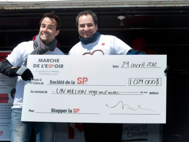
Sbastien Benoit, TV and Radio Host, provincial spokesperson, and Louis Adam, Executive Director, of the MS Society
MONTREAL, April 29, 2012 /CNW Telbec/ - On Sunday, Walkers of all ages turned out for the 18th MS Walk for the benefit of the Multiple Sclerosis Society of Canada. Through their efforts, over $1,029,000 was raised.
The Walk was held in 16 regions of Quebec: Dollard-des-Ormeaux, Blainville, Drummondville, East of Montreal, Gatineau, Granby, Laval, Lvis, Montreal, Mont-Tremblant, Repentigny, Saint-Hyacinthe, Saint-Jrme, Sainte-Catherine, Sept-les and Trois-Rivires. These are preliminary results as we will hold seven other Walks in June and September. Please visit mswalks.ca for further information.
Television and radio host Sbastien Benoit, provincial spokesperson of the MS Walk, joined the event in Montreal for a sixth consecutive year.
The Multiple Sclerosis Society of Canada would like to thank all the Walkers, volunteers and organizers who made this event a huge success!
Multiple sclerosis is the most common neurological disease affecting young adults in Canada and near 20,000 people have MS in Quebec. It affects three times more women than men. Every step matters. Together we can end MS.
End MS
For more information on how to help persons with MS and contribute to the progress of research on the disease, visit http://www.scleroseenplaques.ca/qc or call 514-849-7591 or toll-free in Quebec at 1-800-268-7582.
Image with caption: "Sbastien Benoit, TV and Radio Host, provincial spokesperson, and Louis Adam, Executive Director, of the MS Society - Quebec Division (CNW Group/Multiple Sclerosis Society of Canada )". Image available at: http://photos.newswire.ca/images/download/20120429_C2233_PHOTO_EN_12852.jpg
Source: Marie-Eve Ouellet Communications Coordinator Multiple Sclerosis Society of Canada Quebec Division 514-849-7591 ext. 245 1-800-268-7582 (toll-free in Quebec) ormarie-eve.ouellet@scleroseenplaques.ca.

Read this article:
We've walked for people living with Multiple Sclerosis!
Recommendation and review posted by simmons
Duke team turns scar tissue into heart muscle without using stem cells
Public release date: 26-Apr-2012 [ | E-mail | Share ]
Contact: Sarah Avery sarah.avery@duke.edu 919-660-1306 Duke University Medical Center
DURHAM, N.C. Scientists at Duke University Medical Center have shown the ability to turn scar tissue that forms after a heart attack into heart muscle cells using a new process that eliminates the need for stem cell transplant.
The study, published online April 26 in the journal Circulation Research, used molecules called microRNAs to trigger the cardiac tissue conversion in a lab dish and, for the first time, in a living mouse, demonstrating the potential of a simpler process for tissue regeneration.
If additional studies confirm the approach in human cells, it could lead to a new way for treating many of the 23 million people worldwide who suffer heart failure, which is often caused by scar tissue that develops after a heart attack. The approach could also have benefit beyond heart disease.
"This is a significant finding with many therapeutic implications," said Victor J. Dzau, M.D., a senior author on the study who is James B. Duke professor of medicine and chancellor of health affairs at Duke University. "If you can do this in the heart, you can do it in the brain, the kidneys and other tissues. This is a whole new way of regenerating tissue."
To initiate the regeneration, Dzau's team at Duke used microRNAs, which are molecules that serve as master regulators controlling the activity of multiple genes. Tailored in a specific combination, the microRNAs were delivered into scar tissue cells called fibroblasts, which develop after a heart attack and impair the organ's ability to pump blood.
Once deployed, the microRNAs reprogrammed fibroblasts to become cells resembling the cardiomyocytes that make up heart muscle. The Duke team not only proved this concept in the laboratory, but also demonstrated that the cell conversion could occur inside the body of a mouse a major requirement for regenerative medicine to become a potential therapy.
"This is one of the exciting things about our study," said Maria Mirotsou, PhD, assistant professor of cardiology at Duke and a senior author of the study. "We were able to achieve this tissue conversion in the heart with these microRNAs, which may be more practical for direct delivery into cells and allow for possible development of therapies without using genetic methods or transplantation of stem cells."
The researchers said using microRNA for tissue regeneration has several potential advantages over genetic methods or transplantation of stem cells, which have been difficult to manage inside the body. Notably, the microRNA process eliminates technical problems such as genetic alterations, while also avoiding the ethical dilemmas posed by stem cells.
Excerpt from:
Duke team turns scar tissue into heart muscle without using stem cells
Recommendation and review posted by sam
Woman’s plight spurs push for marrow registry – feature
BEIRUT: A Lebanese expatriate in Australia in need of a bone marrow transplant is looking to find a match in Lebanon and in the process is working to raise awareness to create a national registry of donors in Lebanon.
It would be amazing if there could be a bone marrow registry in Lebanon, says Pamela Bou Sejean. Im getting treatment to keep my cancer under control. But I cant go on too long, said 26-year-old Lebanese expatriate from Western Australia who was diagnosed in 2010 with Hodgkins lymphoma, a type of leukemia that can be treated with a bone marrow transplant.
Bone marrow transplants treat different types of blood cancer, including leukemia, lymphoma and multiple myloma, as well as several non-malignant conditions. The United States and Western Europe have databases for anonymous donors, with Germany having approximately 1 million registered. The best matches tend to be from donors related to the patient, and when that fails, doctors turn to their national registries.
None of Bou Sejeans relatives turned out to be a match, nor was anyone on the national registry in Australia. Her doctors told her that her best hope would be to find a match in her ancestral country of Lebanon, where potential donors have similar genes.
However, until now, bone marrow transplants in Lebanon have almost all been done with relatives of patients because of a lack of a national registry, as well as government funding or charities that can cover the $500 cost of the donor test.
Australia, on the other hand, has been covering the entire cost of Bou Sejeans treatment, including a $10,000 stem cell transplant last year and all of her tests in Lebanon.
Bou Sejean is hoping that her story will help raise enough awareness to push the government to create a national registry program and also encourage Lebanese citizens to take the test and the chance to save someones life.
If a registry is available, people can take the test, Bou Sejean says. Its not painful. Its just like a blood test. It takes the blood and separates the stem cells. To save someones life is worth it.
Bou Sejeans public awareness campaign started when a friend told her local newspaper, the Geelong Advertiser, about her situation. Other media interviews followed. When it was announced that President Michel Sleiman would be traveling to Australia, a newspaper put her in contact with his staff, who arranged a private meeting that lasted half an hour during his state visit.
During a speech in which he addressed members of Lebanons expatriate community in Melbourne, Sleiman implored the Lebanese to take the test to see if they are a match, rhetorically asking, Are we incapable of giving Pamela blood?
Read the original post:
Woman’s plight spurs push for marrow registry - feature
Recommendation and review posted by sam
NIGERIA: Bone marrow register an important milestone
Stem cell transplant wait time considerably longer in poor countries. Nigeria's recently established bone marrow registry promises to boost lifesaving matches
Some 200,000 babies are born annually in sub-Saharan Africa with sickle cell disease, a blood disorder in which mutated red blood cells can clump and block blood vessels, causing pain, infection and organ damage. Nigeria has up to two million sickle cell patients, many of whom can benefit from stem cell transplants.
Stem cells are the building blocks of blood and immune cells. Establishing the mechanics of stem cell transplantation in Nigeria is a very important milestone, said Terry Schlaphoff, deputy director of South Africas bone marrow registry.
Bone marrow registries hold key information about stem cell donors to help match them with patients. There are currently two such registries in Africa, one in South Africa and now Nigeria.
In countries with low per capita incomes, stem cell transplants remain relatively rare due to lack of knowledge, trained health workers and, most importantly, availability of stem cells. African patients who need a matching donor have virtually no chance of survival, unless they are wealthy enough to travel abroad for treatment, said Seun Adebiyi, founder of Nigerias bone marrow registry.
Matching bone marrow or blood cells collected from donors to the patients who need it can offer lifesaving treatments for more than 70 diseases, including leukaemia, lymphoma (cancer) and sickle-cell anaemia.
Limited availability
Worldwide, there are fewer than 15 million registered donors, and patients far outstrip the number of donors, according to the Netherlands-based information centre, Bone Marrow Donors Worldwide (BMDW).
Reflecting only a fraction of overall need, 14,206 transplants from non-relatives and 4,255 transplants from umbilical cord blood were provided to patients worldwide in 2011, said Machteld Oudshoorn, chair of BMDWs editorial board.
For most patients in developing countries, awaiting a transplant remains associated with significant morbidity and mortality, and represents one example of high-cost, highly specialized medicine, according to a recent medical report.
More:
NIGERIA: Bone marrow register an important milestone
Recommendation and review posted by sam
Scientists Reprogram Cells To Heal Broken Hearts
Heart attacks, like any wound, often leave behind scar tissue. But a scarred heart isn't just disfigured, it's weaker -- scar tissue can't contract so the organ loses some of its pumping ability.
If only a fairy could wave a magic wand to transform scar tissue into heart muscle cells.
Well, that transformation has happened in mice -- but the fairy was a team of Duke University Medical Center scientists, and the magic wand was a kind of small molecule called microRNA, which can turn many kinds of genes on or off.
Scientists administered microRNA to scar tissue cells both in petri dishes and in the hearts of living mice, which reprogrammed the scar tissue cells into muscle cells that power the heart; no stem cells or surgery required.
The scientists, led by senior author Victor J. Dzau, reported their findings on Tuesday in the journal Circulation Research.
Scientists need to conduct further studies to examine how the transformed tissue performs compared to the rest of the heart, Dzau said in a telephone interview. And it still remains to be seen if the method works on human hearts.
However, this proof of concept is important in that it shows that scar tissue can be reprogrammed in living animals.
Similar reprogramming may be possible for scar tissues found in other organs including brains and kidneys, Dzau said.
The Duke research is similar to findings reported by Gladstone Institutes scientists in Nature last week. But Gladstone researchers used a different method to reprogram the scar tissue, giving mice a cocktail of three genes instead of microRNA.
The effect was essentially the same. Genes that encouraged the development of heart muscle characteristics were switched on, and the scar tissue gradually metamorphosed into myocardial cells of the heart muscle.
Continued here:
Scientists Reprogram Cells To Heal Broken Hearts
Recommendation and review posted by sam
Lab-on-a-Chip Enables Personalized Medicine – Video
25-04-2012 14:45 A plastic chip can whether a patient is resistant to cancer drugs or has diseases like malaria. The chip can also find infectious diseases in a herd of cattle. Source: Univ. of Alberta Read more:
Read the original here:
Lab-on-a-Chip Enables Personalized Medicine - Video
Recommendation and review posted by sam
Regenerative Medicine Session at Sacramento Med Tech Showcase 2011 – Video
25-04-2012 23:54 Regenerative Medicine session at the 2011 Sacramento Med Tech Showcase by SARTA MedStart
See the original post here:
Regenerative Medicine Session at Sacramento Med Tech Showcase 2011 - Video
Recommendation and review posted by sam
Advanced Cell Technology Announces Webcast of Corporate Presentation at Annual Shareholders’ Meeting Today
MARLBOROUGH, Mass.--(BUSINESS WIRE)--
Advanced Cell Technology, Inc. (ACT;OTCBB: ACTC), a leader in the field of regenerative medicine, announced that it will webcast todays corporate presentation at 9:15 a.m. PDT, after the formal portion of its Annual Shareholders Meeting has concluded. Prior to the webcast, proxy voting will be completed and the results announced to the shareholders in attendance. The meeting is open to shareholders of record as of March 1, 2012. After the corporate presentation, the Company will address questions from shareholders in attendance, as well as electronically via the webcast.
The webcast will be available live and via replay at: http://us.meeting-stream.com/advancedcelltechnology042612
The Presentation schedule is as follows:
About Advanced Cell Technology, Inc.
Advanced Cell Technology, Inc., is a biotechnology company applying cellular technology in the field of regenerative medicine. For more information, visitwww.advancedcell.com.
Forward-Looking Statements
Statements in this news release regarding future financial and operating results, future growth in research and development programs, potential applications of our technology, opportunities for the company and any other statements about the future expectations, beliefs, goals, plans, or prospects expressed by management constitute forward-looking statements within the meaning of the Private Securities Litigation Reform Act of 1995. Any statements that are not statements of historical fact (including statements containing the words will, believes, plans, anticipates, expects, estimates, and similar expressions) should also be considered to be forward-looking statements. There are a number of important factors that could cause actual results or events to differ materially from those indicated by such forward-looking statements, including: limited operating history, need for future capital, risks inherent in the development and commercialization of potential products, protection of our intellectual property, and economic conditions generally. Additional information on potential factors that could affect our results and other risks and uncertainties are detailed from time to time in the companys periodic reports, including the report on Form 10-K for the year ended December 31, 2011. Forward-looking statements are based on the beliefs, opinions, and expectations of the companys management at the time they are made, and the company does not assume any obligation to update its forward-looking statements if those beliefs, opinions, expectations, or other circumstances should change. Forward-looking statements are based on the beliefs, opinions, and expectations of the companys management at the time they are made, and the company does not assume any obligation to update its forward-looking statements if those beliefs, opinions, expectations, or other circumstances should change. There can be no assurance that the Companys clinical trials will be successful.
Original post:
Advanced Cell Technology Announces Webcast of Corporate Presentation at Annual Shareholders’ Meeting Today
Recommendation and review posted by sam
Stem cell researchers map new knowledge about insulin production
Public release date: 26-Apr-2012 [ | E-mail | Share ]
Contact: Professor Palle Serup palle.serup@sund.ku.dk 01-145-402-20026 University of Copenhagen
Scientists from The Danish Stem Cell Center (DanStem) at the University of Copenhagen and Hagedorn Research Institute have gained new insight into the signaling paths that control the body's insulin production. This is important knowledge with respect to their final goal: the conversion of stem cells into insulin-producing beta cells that can be implanted into patients who need them. The research results have just been published in the well-respected journal PNAS.
Insulin is a hormone produced by beta cells in the pancreas. If these beta cells are defective, the body develops diabetes. Insulin is vital to life and therefore today the people who cannot produce their own in sufficient quantities, or at all, receive carefully measured doses often via several daily injections. Scientists hope that in the not-so-distant future it will be possible to treat diabetes more effectively and prevent secondary diseases such as cardiac disease, blindness and nerve and kidney complications by offering diabetes patients implants of new, well-functioning, stem-cell-based beta cells.
"In order to get stem cells to develop into insulin-producing beta cells, it is necessary to know what signaling mechanisms normally control the creation of beta cells during fetal development. This is what our new research results can contribute," explains Professor Palle Serup from DanStem.
"When we know the signaling paths, we can copy them in test tubes and thus in time convert stem cells to beta cells," says Professor Serup.
The new research results were obtained in a cooperative effort between DanStem, the Danish Hagedorn Research Institute and international partners in Japan, Germany, Korea and the USA. The scientific paper has just been published in the well-respected international journal PNAS (Proceedings of the National Academy of Sciences of the United States of America) entitled Mind bomb 1 is required for pancreatic -cell formation.
Better control of stem cells
The signaling mechanism that controls the first steps of the development from stem cells to beta cells has long been known.
"Our research contributes knowledge about the next step in development and the signaling involved in the communication between cells an area that has not been extensively described. This new knowledge about the ability of the so-called Notch signaling first to inhibit and then to stimulate the creation of hormone-producing cells is crucially important to being able to control stem cells better when working with them in test tubes," explains Professor Palle Serup .
More here:
Stem cell researchers map new knowledge about insulin production
Recommendation and review posted by Bethany Smith
Biz Beat: Making stem cells "available to the masses"
Mike Ivey writes on all matters money in the spirit of Capital Times founder William T. Evjue, who believed that the concentration of wealth in the U.S. is not healthy for the Democracy.
When UW-Madison's James Thomson in 1998 became the first scientist to grow human embryonic stem cells in a lab, it generated tremendous excitement about the medical possibilities.
Thomson tried to downplay the breakthrough but talk spread about cures for Alzheimers or Parkinsons disease, growing livers for cirrhosis suffers or producing healthy heart cells for cardiac patients.
The miracle cures have been slow in coming, however. Scientists can replicate healthy nerve cells in a Petri dish but havent found a way to replace defective spinal cells in ALS victims, for example.
In many ways, were still at the first steps,Anita Bhattacharyya, a senior scientist in the stem cell program at the UW's Waisman Center, told a business group Tuesday.
Butproducing stem cells for others to use is proving one of Madisons more promising new business ventures. Pharmaceutical companies in particular are using stem cells to test drugs before launching into expensive further testing.
Were making these cells available to the masses, says Chris Parker, chief technology officer at Cellular Dynamics International.
Launched by Thomson -- and backed with $100 million from a local investor group -- Cellular Dynamics International was lauded recently by MIT as one of the 50 most important companies in the world
Since its founding in 2005, the company now counts 107 employees at it offices in University Research Park and is continuing to grow.
Im hiring right now, Parker joked toa lunch crowd of the Wisconsin Technology Council Tuesday.
Continued here:
Biz Beat: Making stem cells "available to the masses"
Recommendation and review posted by Bethany Smith